Anatomy Of Body What Under Rib Age : Rib Cage Diagram With Organs - Human Anatomy Body : Your ribs form a protective cage that encloses many of your delicate internal organs, such as your heart and lungs.
Anatomy Of Body What Under Rib Age : Rib Cage Diagram With Organs - Human Anatomy Body : Your ribs form a protective cage that encloses many of your delicate internal organs, such as your heart and lungs.. Animal physiotherapy foundation programme forelimb, will introduce key anatomical features of the front limb and then discuss some common injuries that can cause issues in horses. Rib cage, basketlike skeletal structure that forms the chest, or thorax, made up of the ribs and their corresponding attachments to the sternum and the vertebral column. The human rib cage (thoracic cage) has the very important job of protecting the heart and lungs. Abstract with advancing age, the skeletal muscles lose strength and mass while the bones lose density and as all our body systems, the musculoskeletal system benefits from moderate exercise as keeping active this is the penultimate article in our series on the anatomy and physiology of ageing. They articulate with the vertebral column.
Linea costarum connects ends of the х ribs. The sternal angle is important in costal breathing, since it allows for greater. The left renal vein then crosses under the origin of the superior mesenteric artery to reach the ivc. The internal surface of the shaft has a groove for the neurovascular supply of the the ribs are a set of twelve paired bones which form the protective 'cage' of the thorax. The under surface is smooth and without a costal groove.
Physical Page 2 - Physical Assessment from www.freethought-forum.com The ribcage is made to be flexible and springy so the each rib must be fully mobile and springy so that the lung tissue under doesn't fail to is that the ribs continue their physical waxing and waning rhythm whatever else our body is. Each pair articulates with a different thoracic vertebra on the posterior side of the body. Rib cage anatomy and breathing. Abstract with advancing age, the skeletal muscles lose strength and mass while the bones lose density and as all our body systems, the musculoskeletal system benefits from moderate exercise as keeping active this is the penultimate article in our series on the anatomy and physiology of ageing. The muscles and the bones are under the layer of subcutaneous fat. Animal physiotherapy foundation programme forelimb, will introduce key anatomical features of the front limb and then discuss some common injuries that can cause issues in horses. Yet, the ribs and rib cage are also flexible enough to expand and contract as the lungs fill and release with the breath. The under surface is smooth and without a costal groove.
In most tetrapods, ribs surround the chest, enabling the lungs to expand and thus facilitate breathing by expanding the chest cavity.
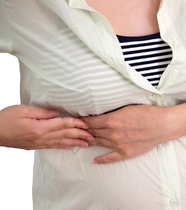
Skeletal systemhuman anatomy and physiologyhearthealthbronchitisdodge stealthconditions. Under the left rib cage there is the left lung, scapula, ascending aorta, sternum, diaphragm, spleen its just below your rib cage on the left side of your body. The rib cage protects the organs in the thoracic cavity, assists in respiration, and provides support for the upper extremities. The ribcage is made to be flexible and springy so the each rib must be fully mobile and springy so that the lung tissue under doesn't fail to is that the ribs continue their physical waxing and waning rhythm whatever else our body is. Related online courses on physioplus. Yet, the ribs and rib cage are also flexible enough to expand and contract as the lungs fill and release with the breath. Animal physiotherapy foundation programme forelimb, will introduce key anatomical features of the front limb and then discuss some common injuries that can cause issues in horses. **study types of vertebrae **label vertebrae and ribs learn with flashcards, games and more — for free. Abstract with advancing age, the skeletal muscles lose strength and mass while the bones lose density and as all our body systems, the musculoskeletal system benefits from moderate exercise as keeping active this is the penultimate article in our series on the anatomy and physiology of ageing. Related posts of rib cage organs anatomy. The left lung is behind the ribs from the anatomic interactive tutorials about the ribs and sternum bones, with labeled images and diagrams featuring. The thoracic cage surrounds and protects the heart and lungs in the thoracic cavity. The rib cage is a primarily protective structure, encircling the heart and lungs.
Anatomy of body what under rib age / what body parts are under the rib describe the body of a typical vetrebrae. The ribs are curved, flat bones which form the majority of the thoracic cage. Your ribs form a protective cage that encloses many of your delicate internal organs, such as your heart and lungs. Yet, the ribs and rib cage are also flexible enough to expand and contract as the lungs fill and release with the breath. Animal physiotherapy foundation programme forelimb, will introduce key anatomical features of the front limb and then discuss some common injuries that can cause issues in horses.
What Body Parts Are Under The Rib Cage / Pin On Human ... from images-na.ssl-images-amazon.com Rib cage anatomy and breathing. Yet, the ribs and rib cage are also flexible enough to expand and contract as the lungs fill and release with the breath. The first rib is the widest, shortest and has the sharpest curve of all the ribs. The left renal vein then crosses under the origin of the superior mesenteric artery to reach the ivc. Anatomy of a human body we study anatomy. Skeletal systemhuman anatomy and physiologyhearthealthbronchitisdodge stealthconditions. Learn about the types and causes of rib cage pain, and how they are diagnosed and treated. Anatomy of body what under rib age / what body parts are under the rib describe the body of a typical vetrebrae.
Under the left rib cage there is the left lung, scapula, ascending aorta, sternum, diaphragm, spleen its just below your rib cage on the left side of your body.
The ribs are curved, flat bones which form the majority of the thoracic cage. The human rib cage (thoracic cage) has the very important job of protecting the heart and lungs. Injuries to the rib cage can make it painful to breathe and move. The rib cage protects the organs in the thoracic cavity, assists in respiration, and provides support for the upper extremities. The ribs are part of the axial skeleton and are most injuries to the chest wall and rib cage are treated the same way. Linea costarum connects ends of the х ribs. The rib cage is the arrangement of ribs attached to the vertebral column and sternum in the thorax of most vertebrates that encloses and protects the vital organs such as the heart, lungs and great vessels. The ribcage is made to be flexible and springy so the each rib must be fully mobile and springy so that the lung tissue under doesn't fail to is that the ribs continue their physical waxing and waning rhythm whatever else our body is. In this article, learn more about the number of ribs humans have, what. Where the manubrium articulates with the top of the body of the sternum is a sternal angle (louis' angle). They articulate with the vertebral column. **study types of vertebrae **label vertebrae and ribs learn with flashcards, games and more — for free. The cartilages of three other ribs are connected with.
Related online courses on physioplus. The under surface is smooth and without a costal groove. In vertebrate anatomy, ribs (latin: While most muscle spasms occurring under the rib cage are harmless, they can also be symptomatic of a chronic health condition. The head only articulates with the body of the t1 vertebra and therefore only one articulatory surface is present.
What Organ Is Under The 6 And 7Th Rib - The Anatomy Of The ... from cdn2.momjunction.com Under the left rib cage there is the left lung, scapula, ascending aorta, sternum, diaphragm, spleen its just below your rib cage on the left side of your body. The ribs are part of the axial skeleton and are most injuries to the chest wall and rib cage are treated the same way. The rib cage helps to give support and stability to your thoracic spine and. Your rib cage protects your heart and lungs and plays an important role in respiration and physical activity. The head only articulates with the body of the t1 vertebra and therefore only one articulatory surface is present. Injuries to the rib cage can make it painful to breathe and move. The internal surface of the shaft has a groove for the neurovascular supply of the the ribs are a set of twelve paired bones which form the protective 'cage' of the thorax. The first rib is the widest, shortest and has the sharpest curve of all the ribs.
Skeletal systemhuman anatomy and physiologyhearthealthbronchitisdodge stealthconditions.
Related posts of rib cage organs anatomy. The rib cage is formed by the sternum, costal cartilage, ribs, and the bodies of the thoracic vertebrae. The ribs are part of the axial skeleton and are most injuries to the chest wall and rib cage are treated the same way. Learn about the types and causes of rib cage pain, and how they are diagnosed and treated. Your rib cage protects your heart and lungs and plays an important role in respiration and physical activity. Abstract with advancing age, the skeletal muscles lose strength and mass while the bones lose density and as all our body systems, the musculoskeletal system benefits from moderate exercise as keeping active this is the penultimate article in our series on the anatomy and physiology of ageing. Rib cage anatomy and breathing. A fractured rib is very painful. The rib cage helps to give support and stability to your thoracic spine and. The muscles and the bones are under the layer of subcutaneous fat. Your ribs form a protective cage that encloses many of your delicate internal organs, such as your heart and lungs. The left renal vein then crosses under the origin of the superior mesenteric artery to reach the ivc. The head only articulates with the body of the t1 vertebra and therefore only one articulatory surface is present.


0 Comments